Anatomy of Lateral wall of Nose
Lateral wall of nose is formed by
- Ethmoid Bone:
Superior Turbinate.
Middle Turbinate.
Uncinate Process. - Inferior Turbinate Bone
- Maxilla:
Frontal Process. - Palatine Bone:
Perpendicular plate. - Lacrimal Bone.
- Nasal Bones.


LATERAL WALL OF NOSE contains three meatus underneath three turbinates: Turbinates are bony projections covered by Erectile mucosa serves to increase the interior surface area.
Inferior Turbinate:
- A separate bone (Not part of ethmoid bone).
- Irregular surface , grooved by vascular channels.
- Articulates with inferior margin of maxillary hiatus (maxillary process) , ethmoid , palatine and lacrimal bone.
- Separate ossification center ~ 5th month of IU life
- Has submucosal cavernous plexus with large sinusoids – under autonomic control (nasal resistance)
- Covered by respiratory epithelium with large no. of goblet cells
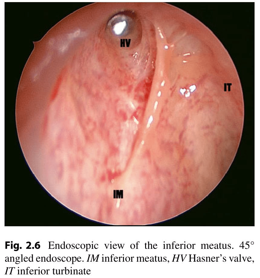
- Inferior meatus: is the area of the lateral wall of the nose covered medially by the inferior turbinate.
- Part of lateral wall of nose lateral to inferior turbinate
- Largest meatus ~ entire length of nasal cavity
- Highest at junction of anterior and middle third.
- 1.6 – 2.3 cm at 1.6 cm
- Nasolacrimal duct opens just anterior to highest point
- Drains: Nasolacrimal Duct (Hasner’s valve).
Middle Turbinate:
- Part of Ethmoid bone.
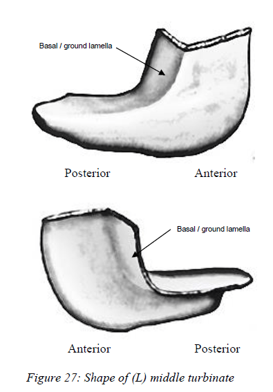
- Attached to Lateral wall by a bony lamella called Ground or Basal Lamella (S-shaped Manner).
- Covered in articulated skull –
- Inferior – maxillary process of IT
- Posterior – perpendicular plate of palatine
- Anterosuperior – portion of lacrimal bone
- Superior – uncinate and bulla of ethmoid.
LATERAL WALL OF NOSE
Middle Turbinate has 3 parts
- Anterior 1/3 attach to cribriform plate and a small anterior attachment to frontonasal process of maxilla
- Middle 1/3 – lamina papyracea
- Posteior 1/3 – lamina papyracea and perpendicular plate of palatine bone
Remaining portion is membranous ( mucosa of middle meatus, maxillary sinus and intervening connective tissue )
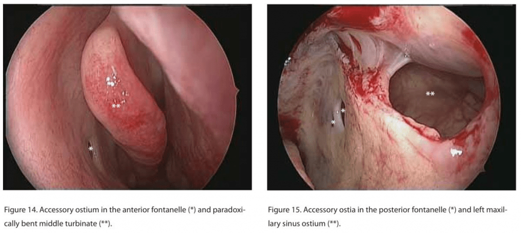
Divided by Uncinate process into anterior and posterior fontanelles
Accessory ostia are found here, Posterior > anterior
AGGER NASI -Small crest or mound on lateral wall just anterior to attachment of middle turbinate. Most anterior part of ethmoid. Occasionally pneumatised (<5% in normal population) – may encroach on NLD •
Basal Lamella of Middle Turbinate
1st Anterior – Sagittal (Vertical) – Skull Base
2nd Middle – Frontal (Oblique) – Lamina Papyracea
3rd Posterior Horizontal – Perpendicular Plate of Palatine Bone

The most anterior part of the middle turbinate fuses with the inferior part of agger nasi to form the “axilla”.
Middle meatus: The area of the lateral wall of the nasal cavity covered medially by the middle turbinate.
Drains: Maxillary sinus, Anterior Ethmoid sinus, Frontal Sinus.
Superior Turbinate:
- Part of ethmoid bone.
- Located above the middle turbinate and bearing olfactory epithelium on its medial surface.
- There may also be a supreme turbinate.
- Superior meatus: The area of the lateral wall of the nose covered medially by the superior turbinate.
Drains:Posterior ethmoid sinuses.
Sphenoethmoidal Recess:
Located in front of the anterior wall of the sphenoid and medial to the superior turbinate. Drains: Sphenoid sinus.

Osteomeatal Complex
Narrow anatomical region in Anterior Ethmoid, Lateral to Middle Turbinate. This is the area bounded by the middle turbiante medially, the lamina papyracea laterally, and the basal lamella superiorly and posteriorly. The inferior and anterior borders of the osteomeatal complex are open.Consist of:
- Bony structures:
o Middle Turbinate.
o Uncinate Process.
o Bulla Ethmoidalis. - Air spaces:
o Frontal Recess.
o Ethmoidal Infundibulum.
o Middle Meatus. - Ostia:
o Anterior Ethmoid Ostium.
o Maxillary Sinus Ostium.
o Frontal Sinus Ostium.

Other DNB ENT Nose Questions
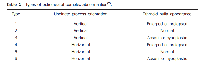
Uncinate Process
Thin, Sickle shaped. First structure encountered in the middle meatus when the middle turbinate is medialized. Forms part of Osteomeatal complex (OMC). It lies in the sagittal plane runs anterosuperior to posteroinferior, with fibrous and bony attachments along the lateral nasal all and forms the medial wall of the ethmoid infundibulum.
Uncinate Process has 3 parts:
- The Horizontal portion:– attached to inferior turbinate ,palatine bone.
- The Middle /Intermediate portion:-attached to lacrimal bone and lamina papyracea .
- The Vertical/Superior portion: -attached to medial orbital wall, middle turbinate and base of the skull base.
SURGICAL IMPORTANCE:-
- First to be removed in various procedures such as MMA leading to infundibulum through haitus semiluminaris.
- Exposes nasolacrimal sac in Endonasal DCR.
- The superior attachment of the uncinate process has implications on the drainage of the frontal sinuses. Determining the frontal recess drainage.
- Preserved during various procedure as it act as guide to frontal sinus in Revision sinus surgeries.
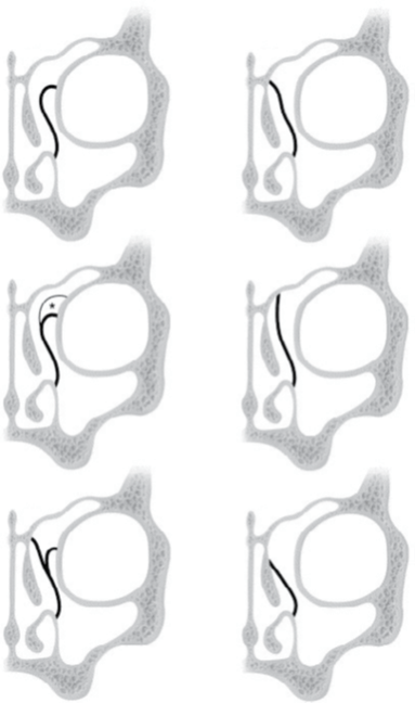
According to Landsberg and Friedman, there are six variations in the attachment of uncinate process superiorly1,2.
•Type 1: Insertion to the lamina papyracea .
•Type 2: Insertion to the posteromedial wall of the agger nasi cell.
•Type 3: Insertion to both the lamina papyracea and the junction of the middle turbinate with the cribriform plate .
•Type 4: Insertion to the junction of the middle turbinate with the cribriform Plate.
•Type 5: Insertion to the skull base.
•Type 6: Insertion to the middle turbinate.
Fontanelles
- Thin boneless segments of lateral nasal wall above the Inferior Turbinate.
- They are closed with mucosa, connective tissue and in continuity with the maxillary periosteum.
- Uncinate Process divides Fontanelles into Anterior and Posterior fontanelle.
- Results in Accessory ostium of Maxillary Sinus, in 5% of the normal population and up to 25% of patients with chronic rhinosinusitis.
- Surgical note: Natural ostium of the maxillary sinus lies between the anterior and posterior fontanelles of the maxillary sinus and cannot usually be seen with a 0 degree endoscope without removing the uncinate process mainly due to its oblique orientation in the sagittal plane. If an ostium is seen, it is most likely an accessory ostium (in the absence of previous sinus surgery).
Agger Nasi:
- Most Anterior ethmoid cell.
- Has a variable degree of pneumatisation in up to 90% of patients.
- It is the first pneumatisation seen on sagittal and coronal CT, posterior to the lacrimal bone and anterior to the free edge of the uncinate process.
- Seen on intranasal examination as a small prominence on the lateral nasal wall:
o Anterior and superior to the attachment of the middle turbinate.
o Anterior to Upper margin of Nasolacrimal duct.
o Posterior wall of Agger nasi cells forms Anterior wall of Frontal recess. - Major dimensions are correlated with Frontal sinus diseases and Lacrimation.
Ethmoidal Infundibulum
- Funnel shaped space.
- Channels drainage of Maxillary, Anterior Ethmoid and Frontal sinuses into Middle Meatus.
- Limited Medially by:
o Uncinate Process.
o Frontal Process of Maxilla. - Limited Laterally by: Lamina Papyracea.
- Limited posteriorly by: Anterior face of ethmoidal bulla.
- Opens into the middle meatus via Hiatus Semilunaris Inferior.
- Natural ostium of Maxillary sinus is situated in Posterior Lower part of Infundibulum.
- Relationship to frontal recess varies with Uncinate Process attachments:
Terminal recess of the ethmoidal infundibulum is formed if the superior attachment of the uncinate process is onto the lamina papyracea or the base of an agger nasi cell, thus forming a blind end to the ethmoidal infundibulum superiorly.

Hiatus Semilunaris Inferior
- Crescent-shaped gap between Posterior free edge of Uncinate Process and Anterior wall of Bulla Ethmoidalis.
- Communication between Middle meatus and Infundibulum.
Hiatus Semilunaris Superior
- Cleft-like communication between Bulla Ethmoidalis and skull base.
- Opens into middle meatus.

Bulla Ethmoidalis
- Most consistent landmark in sinus surgery.
- Largest Anterior Ethmoidal Air cell.
- Situated within Middle meatus
- Posterior to Uncinate Process
- Anterior to Basal Lamella of Middle Turbinate.
- The anterior face of the bulla forms the posterior border of:
o Inferior semilunar hiatus
o Ethmoidal infundibulum
o Frontal recess. - Opens into the superior semilunar hiatus or retrobullar recess (70%).
If the ethmoidal bulla reaches the ethmoidal roof: It forms the posterior border of the frontal recess.
If the ethmoidal bulla does not reaches the ethmoidal roof: An air-filled space called Supra-bullar recess is present between the superior aspect of the bulla and the ethmoidal roof.
Bordered inferiorly by roof of ethmoidal bulla.
Bordered medially by middle turbinate.
Bordered laterally by lamina papyracea.
Bordered superiorly by roof of the ethmoid.
Found in 70% of cadavers.
Laterally it may give rise to an air-containing cleft extending above the orbit, known as a supraorbital recess.
If the posterior wall of ethmoidal bulla is separate from the basal lamella of the middle turbinate: An air-filled space called Retro-bullar recess is present.
Bordered medially by middle turbinate.
Bordered laterally by lamina papyracea.
o It opens medially into the middle meatus via the superior semilunar hiatus.
o Found in 90% of cadavers.
Lateral Sinus: Formed by Supra-bullar + Retro-bullar Recesses. This term has been abandoned.
Conchal Sinus: Space between Middle turbinate and Medial wall of Bulla Ehmoidalis.
Surgical note:
o If the bulla is poorly or non-pneumatized, the medial wall of the orbit is potentially at risk.
o It is also important that the surgeon appreciates the proximity of the skull base when the bulla is pneumatised superiorly.
LATERAL WALL OF NOSE
- 1.Landsberg R, Friedman M. A Computer-Assisted Anatomical Study of the Nasofrontal Region. Laryngoscope. December 2001:2125-2130. doi:10.1097/00005537-200112000-00008
- 2.Güngör G, Okur N, Okur E. Uncinate Process Variations and Their Relationship with Ostiomeatal Complex: A Pictorial Essay of Multidedector Computed Tomography (MDCT) Findings. Pol J Radiol. 2016;81:173-180. doi:10.12659/PJR.895885
LATERAL WALL OF NOSE